Not a fact anymore

Cell membranes are static protein–lipid–protein sandwiches.

What we know now

Cell membranes are flexible double layers of lipid molecules with proteins embedded in or attached to them. The lipids and many proteins can move sideways within the membrane, and cells organize them into changing clusters and specialized regions.

Why it changed

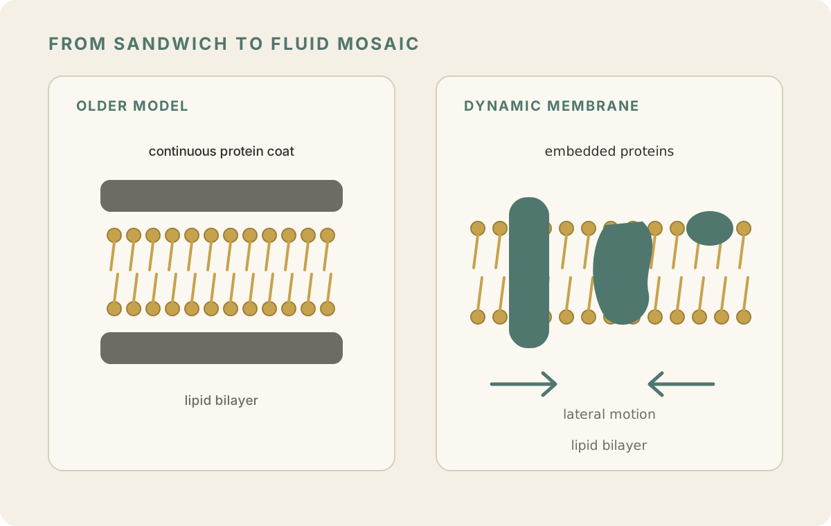
Older models pictured a lipid layer coated on both sides by continuous sheets of protein. Freeze-fracture microscopy instead revealed particles embedded within membranes, and cell-fusion experiments showed membrane proteins moving sideways across the surface. These findings led to the fluid-mosaic model.

Status
Superseded
Category
Cell Biology
Accepted for
≈37 years
Accepted approximately
1930s–1960s
Changed approximately
1960s–1972
Two-panel membrane diagram. The older model shows a lipid bilayer coated on both sides by continuous protein layers. The modernized fluid-mosaic view shows proteins embedded within a lipid bilayer, with arrows indicating lateral movement.

The Danielli–Davson model placed continuous protein coats on both sides of the lipid layer. Later evidence showed many proteins embedded within a fluid bilayer and able to move laterally. Modern membranes are more organized and crowded than this simplified fluid-mosaic sketch.

Original graphic: Not a Fact AnymoreOriginal graphic

Early membrane models tried to explain two things at once: cells clearly had a fatty barrier, but that barrier also had to control what entered and left.

The influential Danielli-Davson model pictured the membrane as a sandwich. A layer of lipids sat in the middle, with continuous layers of protein coating both outer surfaces. Later electron-microscope images seemed to support a neat three-layer structure.

Several experiments made that picture harder to maintain.

Freeze-fracture microscopy splits a frozen membrane apart along its interior. Instead of showing proteins only as surface coatings, it revealed particles embedded within the membrane itself. Biochemical work likewise showed that many membrane proteins extend deeply into the lipid layer.

Cell-fusion experiments supplied another clue. When two cells with differently labeled membrane proteins were fused, the proteins gradually mixed across the combined surface. The membrane was not a rigid sheet; many of its components could move sideways.

Singer and Nicolson brought these findings together in their 1972 fluid-mosaic model: a flexible lipid bilayer with proteins embedded in or associated with it. Modern membranes are even more organized and crowded than that simple picture suggests, but the universal static sandwich did not survive.

Evidence

Sources and what they establish

Previous belief

Primary research

Current evidence

Related entries

Or go somewhere else: